personally performed endoscopic spine operations
The professor's personal cumulative experience in endoscopic spine surgery.
SPINE SURGEON · CLINICAL PRACTICE SINCE 2004
Have screws or a cage been recommended?
Discuss the options for endoscopic and minimally invasive lower-back surgery with Professor Iakiv Fishchenko. Nerve decompression, interbody fusion and percutaneous fixation — with the necessary scope of surgery planned individually.
Preliminary imaging review is free. Contact the professor directly through a messenger.
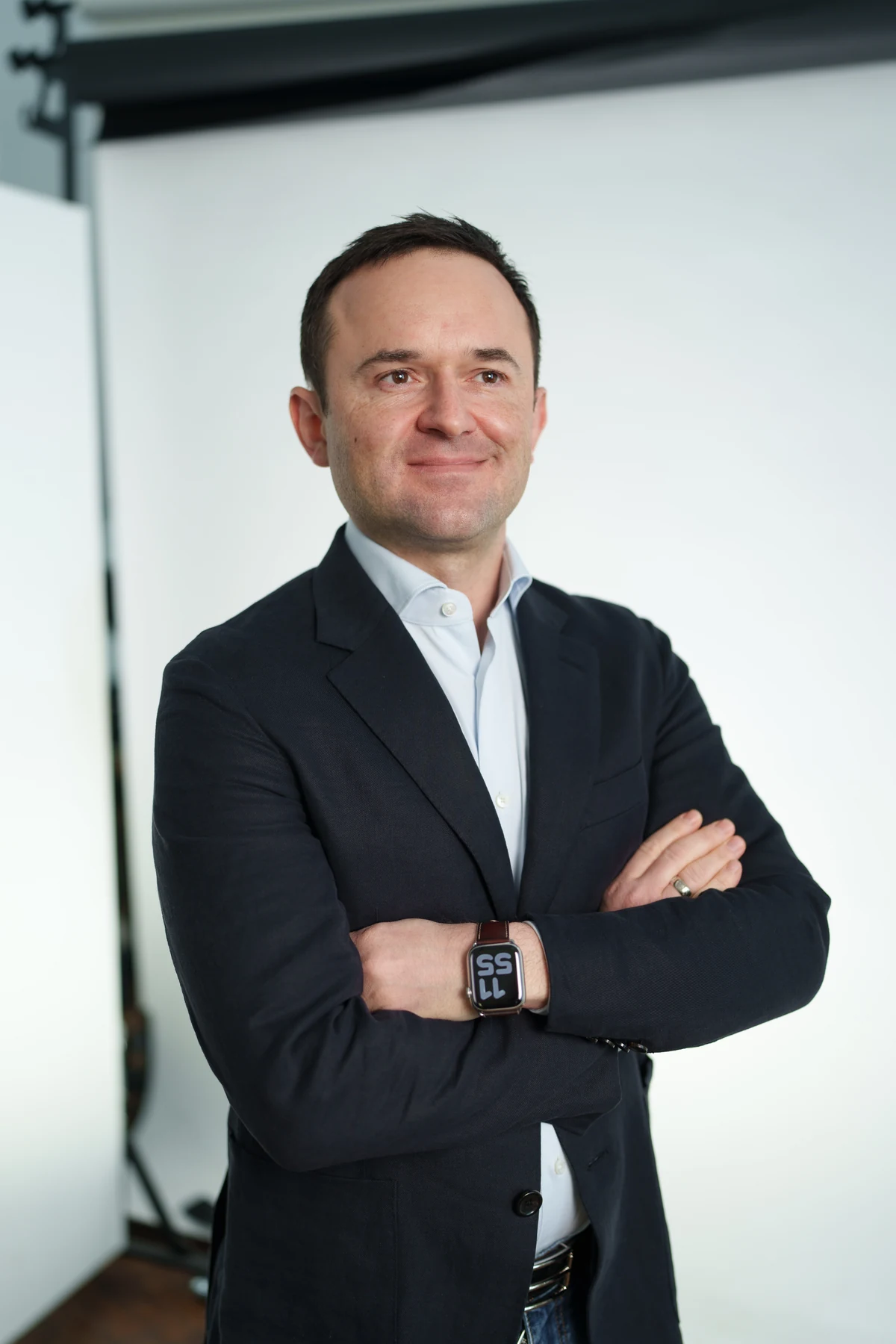
Doctor of Medical Sciences · Spine surgeon
Honoured Doctor of Ukraine · 202601 / PROFESSIONAL EXPERIENCE
Professor Iakiv Fishchenko combines spine surgery, analysis of treatment outcomes and surgical education. Spondylolisthesis, decompression and spinal stabilisation form part of his clinical practice.
The professor's personal cumulative experience in endoscopic spine surgery.
Coauthor of a comparative study of unilateral biportal endoscopic and posterior lumbar interbody fusion.
Author of an interactive atlas of biportal endoscopic spine surgery. Teaching endoscopic surgery since 2019.
02 / UNDERSTANDING THE DIAGNOSIS
Implants are used for different conditions when the spine needs support and stability. The diagnosis is only part of the assessment: the mechanical problem, symptoms, nerves and bone health also matter. A disc herniation or vertebral slippage alone does not mean that screws are needed.
Indications for spinal fusion · AAOS ↗Vertebral slippage and excessive movement: the cause of pain is assessed against imaging and examination findings.
Nerve compression may coexist with mechanical instability. For some patients, decompression alone is sufficient.
Previous treatment or a disturbance of spinal balance may call for reconstruction with fixation.
Certain injuries, infections or tumours require stabilisation alongside treatment of the underlying condition. An endoscopic approach is not suitable for every such case.
03 / TREATMENT PLANNING
A consultation establishes what needs treatment: nerve compression, instability or both. This assessment informs the appropriate scope of surgery.
Where pain occurs, how walking has changed, whether weakness is present and what treatment has already been tried.
MRI shows neural structures. X-rays and, when indicated, flexion–extension views assess vertebral position and movement.
Non-surgical care, decompression and, where indicated, stabilisation with fusion are discussed.
The surgical objective, approach, extent of fixation, risks and recovery are discussed before the operation.
04 / SURGICAL TECHNIQUES
When stabilisation is necessary, Professor Iakiv Fishchenko combines endoscopic decompression of neural structures with interbody fusion and percutaneous pedicle-screw fixation. The cage is inserted exclusively through an endoscopic approach. A cage and bone graft material are always used for interbody fusion.
One construct addresses three objectives: nerve decompression, segmental stability and the formation of bone union. The professor determines whether this operation is needed from your symptoms and investigations.
Endoscopic decompression aims to release compressed nerves.
The cage provides support between the vertebral bodies, while percutaneous screws and rods hold the segment in place. Together they provide reliable stability and distribute the load.
In our interbody fusion technique, a cage and bone graft material are essential components. They provide the conditions for a strong bony union to develop.
The aim is to release compressed nerves. For some patients with degenerative spondylolisthesis, decompression without additional fusion may be sufficient.
More about decompression ↗Screws are inserted through small incisions and connected with rods. In our technique, fixation works together with an interbody cage: the screws hold the segment, while the cage supports it from the front.
Discuss the indications ↗A porous titanium cage is inserted exclusively through an endoscopic approach. It supports disc-space height, distributes the load and, together with bone graft material, provides the conditions for fusion.
Why porous titanium ↗Fixation holds the segment in place, while fusion aims for bone union. These are related but distinct surgical objectives.
A cage is an essential component of stabilisation for degenerative spine disease when we perform endoscopic interbody fusion. It provides support between the vertebrae and complements screw fixation. We use only porous titanium cages, which offer several advantages over plastic PEEK cages.
The porous surface provides the conditions for contact with bone and bone ingrowth into the implant. This supports its biological integration with the vertebrae.
In a clinical comparison, porous titanium cages demonstrated faster early fusion than PEEK cages. The porous structure works together with the bone graft material that we always use for fusion.
A specific porous 3D titanium cage model showed a lower rate of subsidence than PEEK, including in patients with osteoporosis. This advantage supports the choice of porous titanium when bone density is reduced.
Like any interbody cage, it maintains disc-space height and transfers part of the load to the vertebral bodies. Together with screws and rods, this creates stable conditions for fusion.
In osteoporosis, the cage footprint, preservation of the vertebral endplates and treatment of reduced bone density also matter. The properties of the specific implant and the technique used to insert it are considered together.
05 / COMPARING APPROACHES
When stabilisation is needed, the next discussion is how to perform it. A small approach allows focused work while limiting muscle trauma — one of the key advantages of endoscopic surgery.
| What we compare | FOCUS ON TISSUE PRESERVATIONEndoscopic approachInterbody fusion + percutaneous fixation | TRADITIONAL APPROACHOpen surgeryPosterior interbody fusion through an open approach |
|---|---|---|
| Incisions and access | Endoscopic approach Small, separate access points For the camera, instruments and placement of fixation. | Open surgeryA wider surgical approach A longer incision to expose the spinal structures. |
| Muscles and soft tissues | Endoscopic approach Less muscle dissection Limited access reduces trauma to the tissues around the spine. | Open surgeryMore extensive muscle retraction Retraction during an open approach places additional stress on soft tissues. |
| Wound healing | Endoscopic approach Small wounds and tissue preservation Limited damage to the skin and soft tissues creates favourable conditions for healing. This is particularly important in older people and in patients with diabetes or rheumatoid diseases. | Open surgeryA larger wound and more tissue disruption A wider approach leaves a larger surgical wound to heal. Other medical conditions and wound care are taken into account during recovery. |
| View of the surgical area | Endoscopic approach Camera close to the operating site A magnified endoscopic image allows targeted nerve decompression and preparation of the disc space while preserving the bony vertebral endplates. | Open surgeryDirect view through an open approach An operating microscope may be used when needed. |
| Blood loss | Endoscopic approach Lower in several comparative studies A small approach helps limit bleeding. The need for transfusion is assessed individually. | Open surgeryHigher in the same study groups More extensive tissue dissection can increase blood loss. |
| Pain in early recovery | Endoscopic approach Less early back pain This advantage has been shown in comparative studies. Pain relief is still required. | Open surgeryPain from the approach may be more noticeable Muscle trauma adds to recovery from the main stage of surgery. |
| Hospital stay | Endoscopic approach May be shorter This has been reported in some studies; discharge depends on the patient's condition. | Open surgeryMay require more time Recovery from a wider approach can extend the hospital stay. |
| Postoperative scars | Endoscopic approach Several smaller scars Their position and number depend on the levels treated and the planned fixation. | Open surgeryA longer scar Its length reflects the open access required. |
The comparison concerns access to the lumbar spine, not every type of spine surgery. It does not establish a universal timetable for discharge or recovery.
Endoscopic fusion is not necessarily faster: it took longer than open fusion in the cited studies.
Fusion limits movement in the fused segment with either approach. The cited studies did not establish an advantage for endoscopy in fusion rates or overall complication rates.
Open surgery remains appropriate in some complex cases. The choice depends on anatomy, the reconstruction required and the team's experience.
A study involving Professor Fishchenko, Pathologia (2020): 56 patients, a comparison of UBLIF and PLIF, and one-year follow-up. Independent studies published in 2023 and 2024 also assess early recovery after endoscopic and open fusion.
06 / A SCIENTIFIC APPROACH
The professor studies surgical outcomes and contributes to professional discussion. Research helps assess the possibilities and limitations of different techniques.
THE PROFESSOR'S PUBLICATION · PATHOLOGIA · 2020
A study coauthored by Iakiv Fishchenko compared UBLIF and PLIF in 56 patients with lumbar stenosis. It reports one-year follow-up of a specific research cohort.
Read the research paper ↗EVIDENCE-BASED PATIENT SELECTION · BMJ · 2024
Professor Iakiv Fishchenko aims to avoid fusion when decompression can achieve the required result. In the Nordsten-DS trial, decompression alone was non-inferior to the combined operation at five years in selected patients with degenerative spondylolisthesis. The decision is based on clear criteria: symptoms, instability, deformity and the required extent of decompression.
Read Nordsten-DS ↗Lectures, an original atlas and surgical training form part of Iakiv Fishchenko's professional work. SpinaNetUa and ORCID provide access to educational materials and his scientific profile.
07 / PREPARATION
Send complete MRI or CT files with every image series, the report and a brief account of your symptoms. Include existing X-rays and records of previous operations. Further investigations are determined after assessment.
Complete MRI / CT files and existing X-rays.
Symptoms, their duration and previous treatment.
Message the professor on Telegram, Viber or WhatsApp.
The anaesthetist assesses other medical conditions, allergies and medicines. The team determines the necessary tests and investigations, and agrees on the date, treatment location and estimate. Medication plans and fasting instructions before anaesthesia are agreed individually.
Monitoring, pain control, wound care and getting out of bed with the help of a rehabilitation specialist. The timing of mobilisation and discharge depends on the extent of surgery and how you are feeling.
Gradually increasing walking, rehabilitation, follow-up appointments and assessment of bone fusion. Return to work, driving and sport is agreed with the surgeon and rehabilitation specialist.
08 / PATIENT QUESTIONS
The professor's expertise, treatment choices and preparing for consultation.
Professor Iakiv Fishchenko is a spine surgeon and Doctor of Medical Sciences. His practice includes endoscopic decompression, endoscopic interbody fusion and percutaneous screw fixation when indicated. He consults and operates in Kyiv at the National Institute of Traumatology and Orthopaedics of the National Academy of Medical Sciences of Ukraine and the private MedProfi clinic.
Contacts and useful links →Iakiv Fishchenko has been in clinical practice since 2004. Since 2018, he has personally performed more than 3,000 endoscopic spine operations in total. He coauthored a study on interbody fusion, wrote an atlas of biportal endoscopy used to train spine surgeons around the world, and has taught endoscopic spine surgery since 2019.
Slippage on an image alone does not determine treatment. Symptoms, neural findings and previous treatment are assessed. Non-surgical options are discussed at consultation.
No. Decompression may be sufficient for some patients with degenerative spondylolisthesis. The need for fixation is assessed individually.
It is an interbody fusion technique performed through an endoscopic approach: neural structures are decompressed, the disc space is prepared under camera guidance, a cage and bone graft material are inserted, and percutaneous screws complete the construct. A study involving the professor describes the biportal UBLIF technique. Compared with an open approach, it offers several significant advantages: less muscle trauma, less blood loss and less early pain.
Yes. Send current investigations and the records of your previous operation. Professor Fishchenko analyses why the expected result was not achieved, identifies the cause of persistent symptoms and considers possible next steps. The need for another operation is assessed from the investigation findings.
Contacts and useful links →Send complete imaging, a description of your symptoms and the proposed treatment plan to the professor on Telegram, Viber or WhatsApp. Consultation can address the objectives, extent and alternatives to the procedure.
Contacts and useful links →Cost depends on the scope of surgery, implants and treatment location. Recovery is discussed in relation to the planned procedure and your condition. The treatment plan and estimate are clarified with the administrator after consultation.
His scientific profile is available on ORCID. A selected interbody fusion paper is linked in the Research section of this page. Lectures and videos are available on the SpinaNetUa YouTube channel. His full professional profile and atlas of biportal endoscopy are available through the links below.
Contact the professor on Telegram @ProfFishchenko or on Viber / WhatsApp at +380503808725. Information and in-person appointments: 094-821-08-30. Preliminary review helps plan the next step; a final decision requires clinical assessment.
Contacts and useful links →TPF is the Ukrainian abbreviation for pedicle-screw fixation: screws are inserted through the vertebral pedicles and connected with rods. In our interbody fusion technique, a cage and bone graft material are always added. The cage provides support between the vertebral bodies and distributes the load together with the screws. Inadequate fusion and overloading of the construct can lead to implant loosening or damage.
Yes. When stabilisation is necessary, the professor combines endoscopic decompression with endoscopic cage insertion and percutaneous screw fixation. Decompression releases neural structures, the cage and screws stabilise the segment, and bone graft material provides the conditions for fusion. This approach is described in the professor’s patient guide.
In the endoscopic interbody fusion performed by Professor Fishchenko, a cage is an essential component of the construct. It maintains the space between vertebrae, provides support and, together with bone graft material, creates the conditions for fusion. We use only porous titanium: its structure supports bone ingrowth; specific models have shown less subsidence than PEEK, including in osteoporosis.
We use quality branded implants from leading international manufacturers and exclusively porous titanium cages. The professor considers anatomy, bone quality, mechanical loads and the extent of fixation to select the appropriate system, implant size and shape.
Provide the anaesthetist with a complete medication list and information about allergies and chronic conditions. It is particularly important to report anticoagulants and antiplatelet medicines. Do not change them yourself: the doctors agree on a plan based on their indications and risks. Required tests and fasting instructions before anaesthesia are determined individually.
A brace is not needed by every patient. Whether it is necessary and how long to use it depend on the operation and the condition of the spine. The surgeon makes this decision; exercises and activity levels are also agreed with the rehabilitation team.
With an uncomplicated recovery, short journeys as a passenger are usually possible around one week after surgery, and longer journeys around two weeks, after agreement with the professor. Longer trips require breaks for walking. Driving, air travel and return to work or sport are discussed separately, taking account of healing, pain control and safe movement; activity is increased gradually.
Routine removal is not planned after every fusion. It may be considered for specific indications, such as infection or a problem with the construct. The decision takes account of symptoms, imaging and the state of bone fusion.
Yes. We use implants that allow MRI under the manufacturer’s specified conditions. Before the scan, show your discharge summary or implant identification documents to the MRI team so they can check the conditions for the specific system and scanner.
Fusion eliminates movement in the segments intended to unite. With pronounced degenerative changes, these segments often already have limited movement before surgery, so many patients do not notice a substantial additional restriction in everyday life. The effect depends on the number and level of the segments and the condition of the rest of the spine; expected changes and rehabilitation are discussed before surgery.
The main difference is the approach. The camera and instruments work through small openings, the cage is inserted endoscopically and the screws percutaneously. This limits muscle trauma and offers significant advantages in blood loss and early pain. Small wounds create favourable conditions for healing. However, fusion creates bone union with either approach, and the risks of surgery remain.
A small approach preserves soft tissues, reduces blood loss and early postoperative pain, and creates the conditions for early mobilisation. According to Professor Fishchenko’s assessment, an endoscopic operation can be performed in 97% of cases of degenerative lumbar disease that require stabilisation. The duration of surgery and the pace of rehabilitation depend on the extent of the operation and the patient’s condition.
09 / CONSULTATION
Have screws or a cage been proposed? Send your imaging to Professor Iakiv Fishchenko to discuss the indications and options for endoscopic and minimally invasive lumbar stabilisation. The online consultation is completely free.
Complete imaging is sent directly to the professor in your chosen messenger.
Direct contact with Professor Fishchenko
Information and appointments · Kyiv
094-821-08-30fishchenko@gmail.comF0948210830@gmail.com